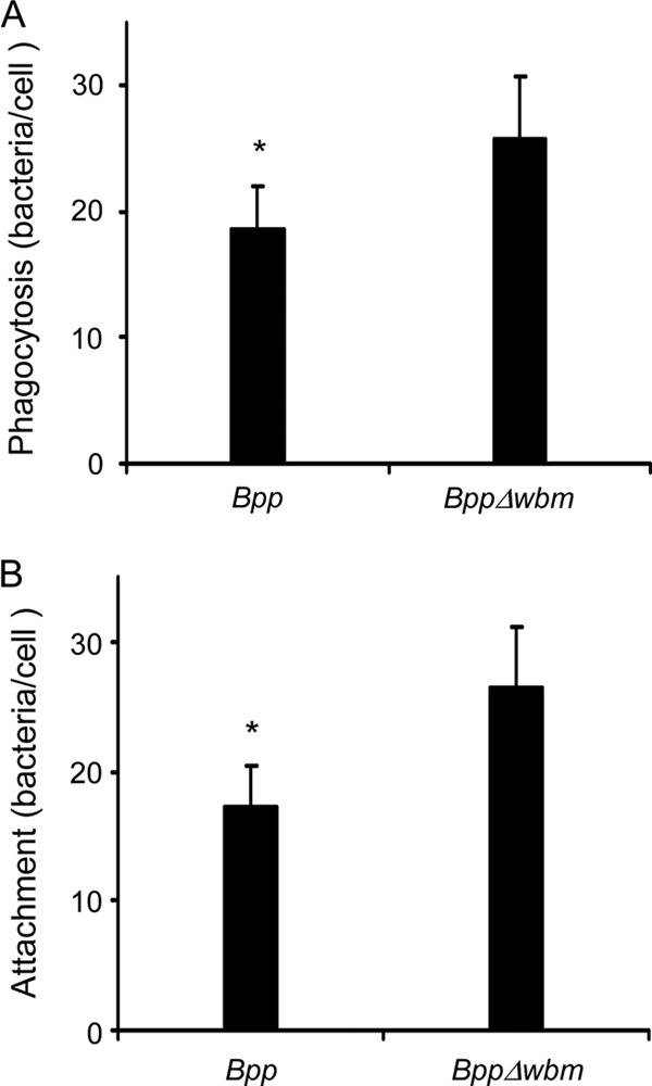
Fig 1.

PMN attachment and phagocytosis of Bordetella parapertussis. (A) PMN phagocytosis of wild-type B. parapertussis (Bpp) and O antigen-deficient B. parapertussis (BppΔwbm). The bacteria were incubated with PMNs (MOI, 300) for 15 min at 37°C. After attachment, the PMNs were washed and further incubated for 1 h at 37°C to allow internalization. The cells were fixed and permeabilized prior to labeling the intracellular bacteria with green fluorescent dye and the extracellular bacteria with both green and red fluorescent dyes. Bacterial phagocytosis was assessed by confocal laser scan fluorescence microscopy. To assess the number of phagocytosed bacteria, at least 100 cells were counted per slide. The data represent the means ± SD of four experiments with PMNs from different donors. Phagocytosis of nonopsonized B. parapertussis by PMNs differed significantly from the PMN phagocytosis of B. parapertussis Δwbm. The asterisk indicates a P value of <0.05. (B) PMN attachment of B. parapertussis. Wild-type B. parapertussis and O antigen-deficient B. parapertussis were incubated with PMNs (MOI, 300) in the presence of cytochalasin D for 15 min at 37°C. PMNs were then washed and further incubated for 1 h at 37°C in the presence of cytochalasin D. The cells were fixed and permeabilized prior to labeling the intracellular bacteria in green fluorescent dye and the extracellular bacteria with both green and red fluorescent dyes. Bacterial attachment was assessed by confocal laser scan fluorescence microscopy. To assess the number of PMN-attached bacteria, at least 100 cells per slide were counted. No bacteria exhibiting only green fluorescence were observed in any cells after 1 h of incubation at 37°C, indicating that phagocytosis was efficiently blocked. The data represent the means ± SD of four experiments with PMNs from different donors. The attachment of B. parapertussis by PMNs differed significantly from PMN attachment of B. parapertussis Δwbm. The asterisk indicates a P value of <0.05.
