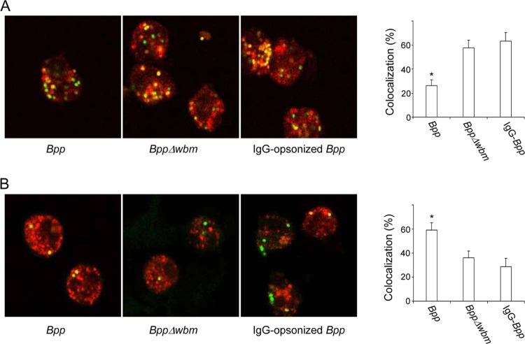
Fig 3.
Confocal laser scanning fluorescence microscopic analyses of B. parapertussis colocalization with LAMP-1 and transferrin. Nonopsonized B. parapertussis (Bpp), nonopsonized B. parapertussis Δwbm, or IgG-opsonized B. parapertussis was incubated with human PMNs (MOI, 300 for nonopsonized bacteria and 30 for IgG-opsonized bacteria) for 15 min at 37°C. After washing, the bacterially infected PMNs were incubated for 1 hour more at 37°C and fixed and permeabilized prior to incubation with antibodies against LAMP-1 (A) or incubated with Alexa transferrin-594 before fixing (B). Shown are green fluorescent bacteria inside PMNs. Colocalization is reflected by the yellow areas. The bars indicate the respective percentages of LAMP-1-positive or transferrin-positive phagosomes. The data represent the means ± SD of three independent experiments. Representative confocal microscopy images of one of three independent experiments are shown. The asterisks indicate significance (P < 0.05).

