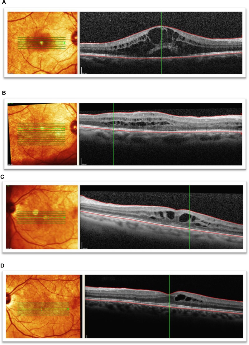
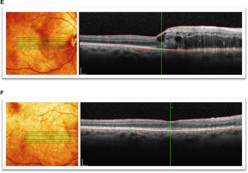
Figure 3.
Spectral domain optical coherence tomographic images of three patients showing improvement in best corrected visual acuity to 20/50 or better following bevacizumab treatments for radiation maculopathy. (A) Patient 1: underwent plaque brachytherapy (right eye) with 20/25 vision at the time. Four years after treatment, the patient presented with subretinal fluid and foveal cysts, grade 6 with best corrected visual acuity of 20/80, and mean foveal thickness of 802 μm. (B) Patient 1: after treatment with six intravitreal bevacizumab treatments over 19 months, visual acuity improved to 20/25 and macular edema improved to grade 4. Mean foveal thickness decreased to 339 μm. (C) Patient 2: underwent plaque brachytherapy (left eye) with 20/400 vision at the time. Twenty-two months after treatment with plaque brachytherapy, the patient presented with grade 5 macular edema and best corrected visual acuity of 20/70 with mean foveal thickness of 483 μm. (D) Patient 2: after treatment with five intravitreal antivascular endothelial growth factor injections over 12 months, visual acuity improved to 20/25, macular edema improved to grade 4, and mean foveal thickness decreased to 366 μm. (E) Patient 3: presented with radiation maculopathy (grade 6) 31 months after treatment with plaque brachytherapy with 20/30 in the right eye and mean foveal thickness of 472 μm. (F) Patient 3: after receiving two intravitreal antivascular endothelial growth factor injections over 10 months, vision improved to 20/30, macular edema grade resolved to normal from grade 6, and mean foveal thickness decreased to 284 μm.


