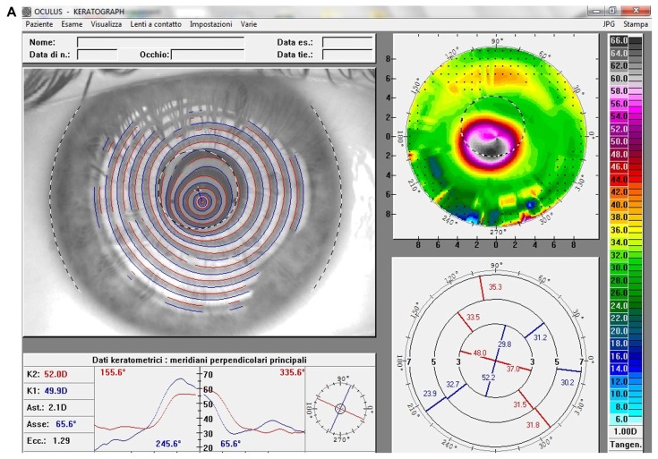
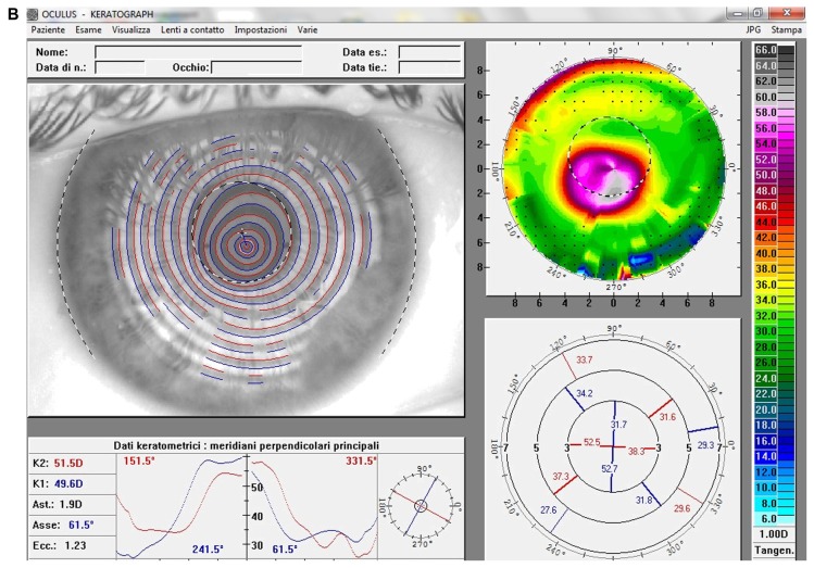
Figure 2.
(A) Videokeratographic map of the right eye of a 19-year-old male patient before treatment. The topographic pattern highlights the keratoconus appearance. The apex of ectasia power (in the central side of the cornea) is 68.7 D (relative scale, tangential algorithm). (B) Right eye videokeratographic map 12 months after transepithelial corneal collagen cross-linking with riboflavin and ultraviolet A irradiation.
Note: The topographic pattern showed a slight improvement in corneal profile, and a reduction of the apex of ectasia power to 65.2 D (relative scale, tangential algorithm).


