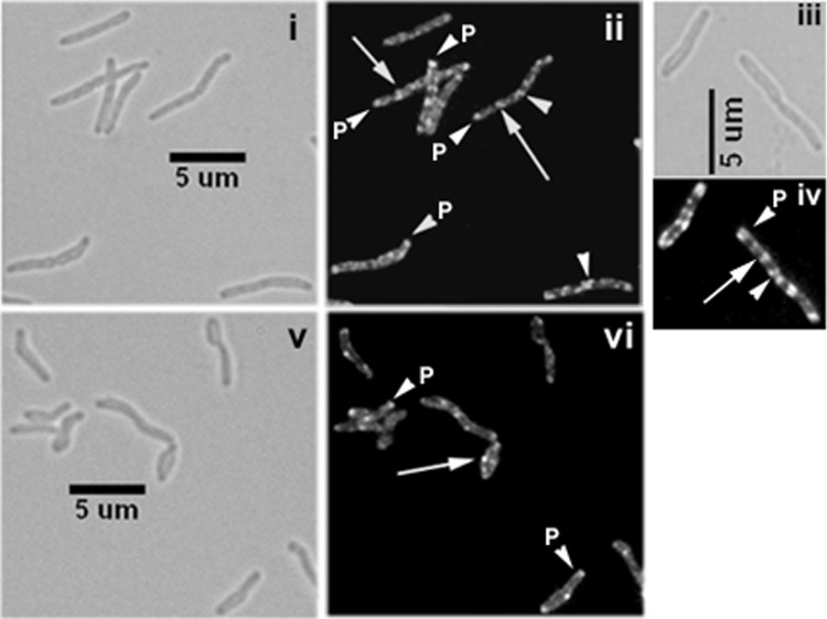
Fig 2.
Cellular localization of CwsA. Exponential cultures of M. smegmatis Pami::gfp-cwsA (i to iv) and M. smegmatis ΔcrgA Pami::gfp-cwsA (v, vi) were induced with 0.2% acetamide for 5 h and visualized by bright-field (i, iii, and v) and fluorescence (ii, iv, and vi) microscopy. Images in panels iii and iv were slightly enlarged to show details of GFP-CwsA localization. White arrows, punctate localization; arrowheads, polar (P) or new pole (midcell) localization.

