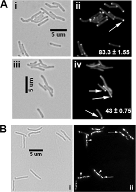
Fig 6.

CwsAsol does not fully complement the defect in the ΔcwsA strain. (A) Exponential cultures of M. smegmatis WT (i, ii) and the ΔcwsA strain complemented with cwsAsol (iii, iv) expressing Pami::wag31-mCherry were induced with 0.2% acetamide for 2 h and imaged by bright-field (i, iii) and fluorescent (ii, iv) microscopy. Arrows, polar localization of Wag31-mCherry. Percent polar localizations from two independent experiments were scored, and averages ± standard errors are given in the respective fluorescent panel. Each experiment included ≥100 cells from each strain. (B) GFP-CwsAsol localization. The M. smegmatis Pami::gfp-cwsAsol strain was grown with acetamide for 5 h and imaged as described in the legend to Fig. 2. (i) Bright-field image; (ii) fluorescent image. Note the polar (arrow) and punctate (arrowhead) patterns of GFP-CwsAsol and absence of membrane localization in panel ii (compare with Fig. 2).
