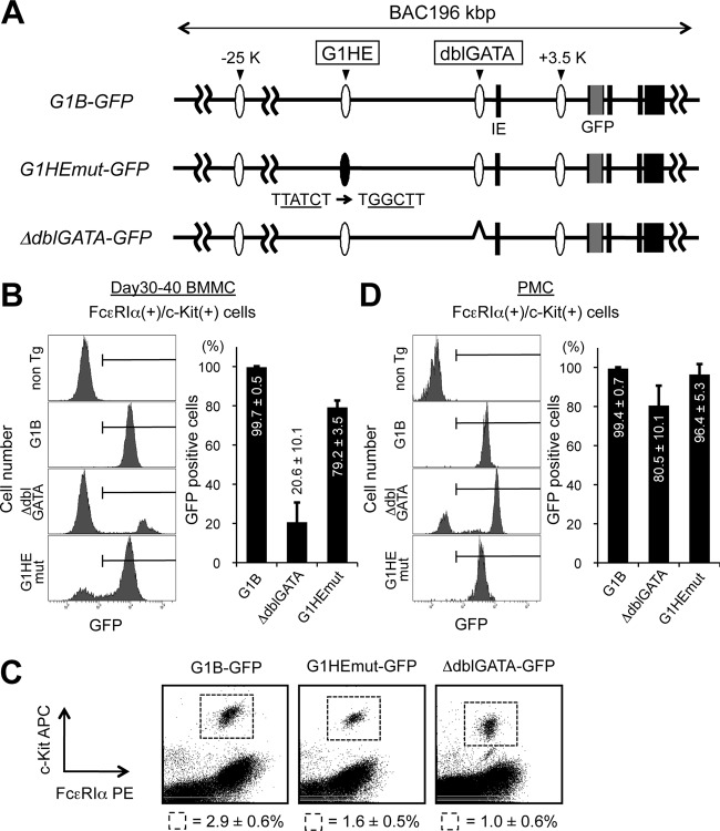
Fig 8.
The G1HE region is dispensable for Gata1 expression in BMMCs. (A) Structures of the G1B-GFP transgenes, namely, G1B-GFP, G1HEmut-GFP, and ΔdblGATA-GFP. The black boxes depict Gata1 gene exons. IE denotes the erythroid cell-specific first exon. GFP cDNA and the conserved GATA sites G1HE and dblGATA are represented by gray boxes and ovals, respectively. (B and D) GFP expression in cultured BMMCs (B) and PMC (D) prepared from G1B-GFP transgenic mice (G1B, G1HEmut, and ΔdblGATA) and nontransgenic control mice (non Tg). The bar graphs show the percentages of cells that were GFP positive within the c-Kit/FcεRIα-double-positive fraction. The results are shown as averages and SD of data obtained from 3 independent experiments. (C) Flow cytometric analysis of peritoneal mast cells isolated from GFP reporter mice stained for c-Kit and FcεRIα expression. The numbers represent the average percentages and SD of c-Kit/FcεRIα-double-positive cells within the indicated gates. The data were obtained from 3 mice for each transgenic line.

