Figure 1.
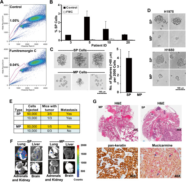
SP cells exhibit stem-like properties. (A) FACS analysis on single cell suspension of human NSCLC xenograft stained with Hoechst 33342 dye showing SP cells. Fumitremorgin C (FTC) inhibited the efflux of the dye and caused the disappearance of SP cells. (B) SP cell frequency in presence or absence of FTC in four different human tumor xenografts. (C) Sphere formation assay on SP or MP cells grown in stem cell selective media for 10 days. The pictures of representative spheres are presented. The bar diagram show the average (±SD) number of spheres formed from 2000 cells. (D) SP or MP cells from H1650 and H1975 cell lines were plated in serum free medium supplemented with EGF and bFGF for 10 days under self-renewal assay conditions. The pictures of representative spheres are depicted (E) Indicated number of SP and MP cells from A549-Luc cell line were implanted into the right lung tissue of SCID mice. The tumor incidence and metastasis was monitored for 12 weeks by bioluminescence imaging. (F) Ex-vivo images of the lung, liver, kidney/adrenals and brain captured at the end of experiment. Metastasis was prevalent in SP cells implanted mice. (G) H&E, pan-keratin (brown staining) and mucicarmine (pink staining, indicated by arrow head) staining of whole lungs from mice implanted with SP or MP cells.

