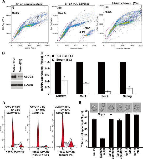
Figure 4.
Establishment and characterization of H1650-SPAdh cells. (A) SP cells from H1650 cell line was plated on normal tissue culture plate (i) or poly-D-lysine-laminin (PDL-Laminin) coated surface (ii) in serum free medium containing N2-supplement, EGF and bFGF. H1650-SPAdh cells growing in self-renewing condition was cultured in serum for 5 days to induce differentiation, and reanalyzed for SP frequency (iii). (B) Serum induces differentiation of SPAdh cells as seen by ABCG2 expression and (C) real time qPCR analysis for stem cell markers, ABCG2, Oct4, Sox2, Nanog. (D) Cell cycle analysis for parental-H1650 or H1650-SPAdh and serum differentiated H1650-SPAdh cells grown on PDL-Laminin coated surface. Histograms were plotted using ModFit program. (E) The average number of spheres generated from 1000 H1650-SPAdh cells is plotted (mean ± SD). Phase contrast microscopy images of the spheres in presence or absence of indicated drugs.

