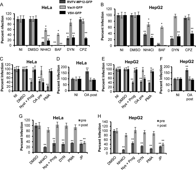
Fig 2.
Inhibitors of caveolar but not clathrin-mediated endocytosis block RVFV infection. Average levels of infection detected by GFP fluorescence (± standard deviations), compared to those for untreated or DMSO-treated controls, are shown. (A and B) HeLa (A) or HepG2 (B) cells were either left untreated (NI) or pretreated with DMSO, 50 mM NH4Cl, 100 nM BAF, 6.5 μg/ml CPZ, or 80 μM DYN for 1 h. The pretreated cells were incubated with the indicated virus at an MOI of 1 for 3 h in the presence of inhibitors. (C and E) HeLa (C) or HepG2 (E) cells were either left untreated or pretreated with DMSO, 30 μM Nys plus 10 μM Prog, 100 nM OA (pre), or 10 μM PMA for 1 h. Inhibitors were also present during 3 h of incubation with RVFV-MP-12-GFP, VacV-GFP, or VSV-GFP at an MOI of 1. (D and F) HeLa (D) or HepG2 (F) cells were infected with the indicated virus at an MOI of 1 for 2 h, washed, and then treated with 100 nM OA for 1 h (post). (G and H) HeLa (G) or HepG2 (H) cells were either left untreated or pretreated (pre) with DMSO, 50 mM NH4Cl, 30 μM Nys plus 10 μM Prog, 80 μM DYN, 10 μM PMA, or 1 μM JP for 1 h. The inhibitors were also present during the 3 h of incubation with RVFV-MP-12-GFP at an MOI of 1. Alternatively, HeLa (G) or HepG2 (H) cells were incubated for 1 h with RVFV-MP-12-GFP at an MOI of 1. Cells were washed to remove unbound virus and were then incubated with the indicated inhibitors (post) for 4 h. GFP expression was normalized to cell titers measured by alamarBlue fluorescence. The percentage of infection was determined by taking untreated or DMSO-treated and infected samples as 100% infected. Shown are the means for three independent experiments performed in triplicate (**, P < 0.01; *, P < 0.05).

