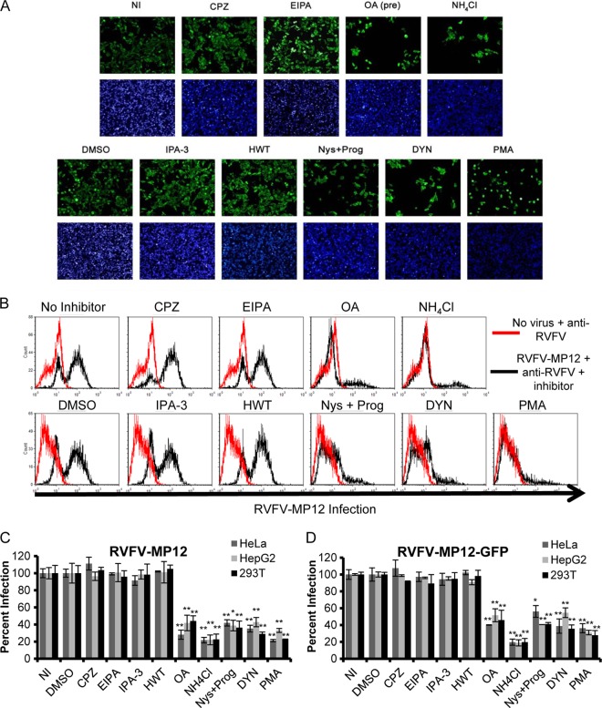
Fig 3.
Inhibitors of caveola-mediated endocytosis, but not inhibitors of CME or macropinocytosis, reduce the percentage of cells infected with authentic RVFV MP-12 or recombinant RVFV-MP-12-GFP. HeLa cells (A, C, and D), 293T cells (B, C, and D), or HepG2 cells (C and D) were either left untreated (NI), pretreated with 6.5 μg/ml CPZ, 25 μM EIPA, 100 nM OA, or 50 mM NH4Cl for 1 h (A and B, top), or treated with DMSO, 10 μM IPA-3, 0.5 μM HWT, 30 μM Nys plus 10 μM Prog, 80 μM DYN, or 10 μM PMA for 1 h (A and B, bottom). The inhibitors were also present during the 3 h of incubation with authentic RVFV MP-12 (A, B, and C) or RVFV-MP-12-GFP (D) at an MOI of 1. (A) Infection of HeLa cells was detected by immunofluorescence using anti-RVFV polyclonal antibodies (green) and the nuclear dye DAPI (blue). This assay was performed in triplicate three or more times with similar results. Images shown are representative of 10 images/well of a 96-well plate. (B) Infection of 293T cells was detected by flow cytometry using anti-RVFV. The data shown are representative of results from three similar experiments performed in duplicate. Red histograms represent uninfected cells that were left untreated (top) or were treated with DMSO (bottom); black histograms represent RVFV-MP-12-infected cells treated with the indicated inhibitors. (C and D) Infection was detected by flow cytometry using an anti-RVFV antibody (C) or GFP expression (D). The quantity of infected cells relative to that of untreated or DMSO-treated controls is given as the percentage of infection. Shown are the means (± standard deviations) for three independent experiments performed in duplicate for each cell type (**, P < 0.01; *, P < 0.05).

