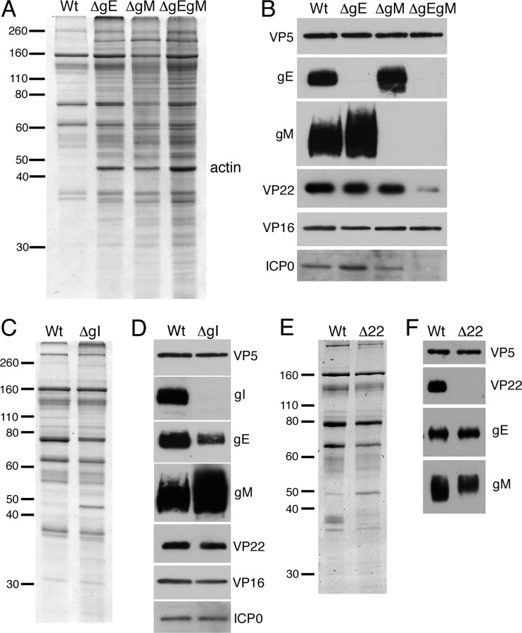
Fig 6.
Relative assembly of VP22 into HSV-1 virions isolated from Vero cells infected with glycoprotein mutant viruses. (A and B) Gradient purified extracellular sc16 (WT), ΔgE, ΔgM, or ΔgEgM virions were analyzed by Coomassie blue staining (A) or by Western blotting using antibodies as indicated (B). Molecular weight marker sizes (kDa) are shown on the left. (C and D) Gradient-purified extracellular sc16 (WT) or ΔgI virions were analyzed by SDS-PAGE followed by Coomassie blue staining (C) or Western blotting with antibodies as indicated (D). Molecular weight marker sizes (kDa) are shown on the left. (E and F) Extracellular WT (s17) or Δ22 virions purified from BHK cells were analyzed by Coomassie blue staining (E) or Western blotting using antibodies as indicated (F). Molecular weight marker sizes (kDa) are shown on the left.

