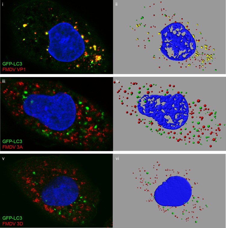
Fig 7.
LC3 punctae generated during FMDV infection contain VP1, but not 3A or 3D. CHO GFP-LC3 cells were infected with FMDV O1BFS (MOI = 2) for 2.5 h in the presence of 10 μM nocodazole. The cells were fixed and immunostained for VP1 (i) (red), 3A (iii) (red), or 3D (v) (red). LC3 was visualized using the natural fluorescence of GFP (green). The panels show side-by-side comparisons of confocal images (i, iii, and v) and digital rendering of pixel densities in fluorescent punctae (ii, iv, and vi) for the same cells.

