Fig 8.
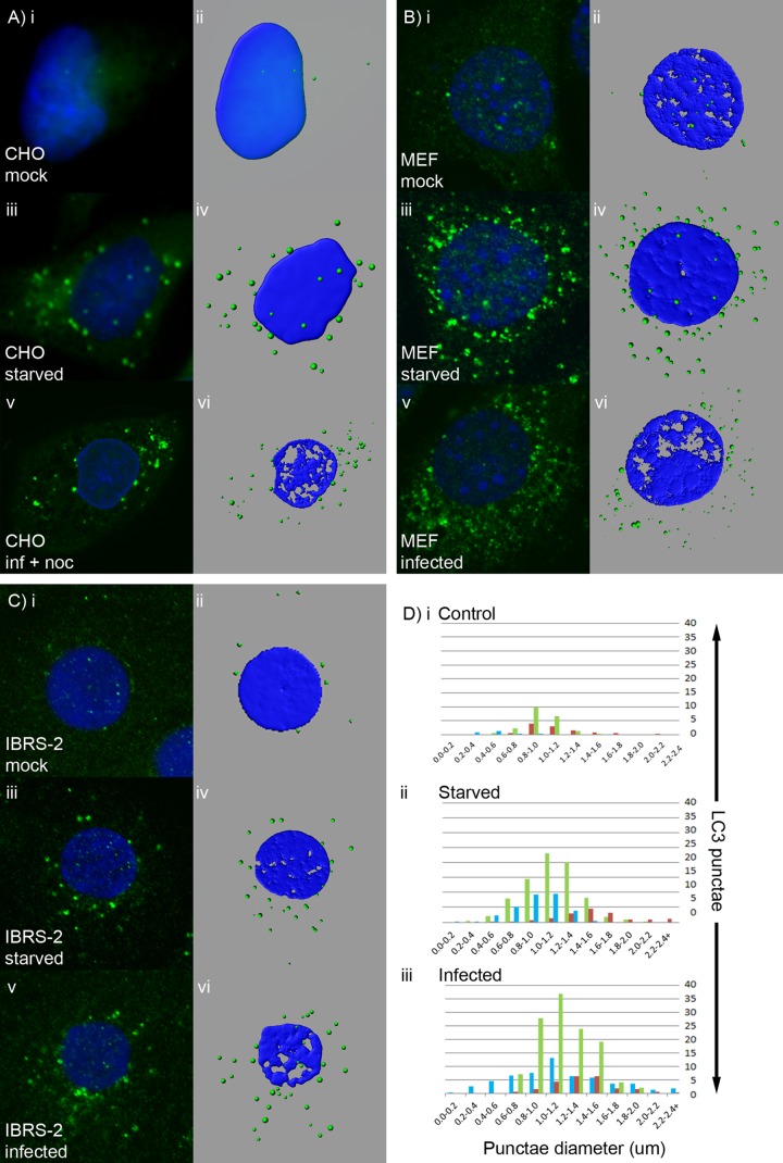
LC3 punctae generated during FMDV infection closely resemble autophagosomes generated in response to starvation. CHO GFP-LC3 cells (A), MEFs (B), or IBRS-2 cells (C) were either mock treated, starved for 2 h, or infected with FMDV O1BFS (MOI = 2) for 2 h (MEFs) or 2.5 h (CHO GFP LC3 and IBRS-2 cells). CHO GFP-LC3 cells were infected in the presence of nocodazole. GFP-LC3 (green) was visualized using the natural fluorescence of GFP. For MEFs and IBRS-2 cells, autophagosome production was analyzed by immunostaining for endogenous LC3 (green). (A to C) Side-by-side comparisons of confocal images (i, iii, and v) and digital rendering of pixel densities in fluorescent punctae (ii, iv, and vi) for the same cells either in control nutrient medium (i and ii), following starvation (iii and iv), or following infection (v and vi). (D) Average numbers of punctae and their diameters calculated from analysis of at least 5 cells (green, MEFs; blue, CHO cells; red, IBRS-2 cells).

