
Fig 1.
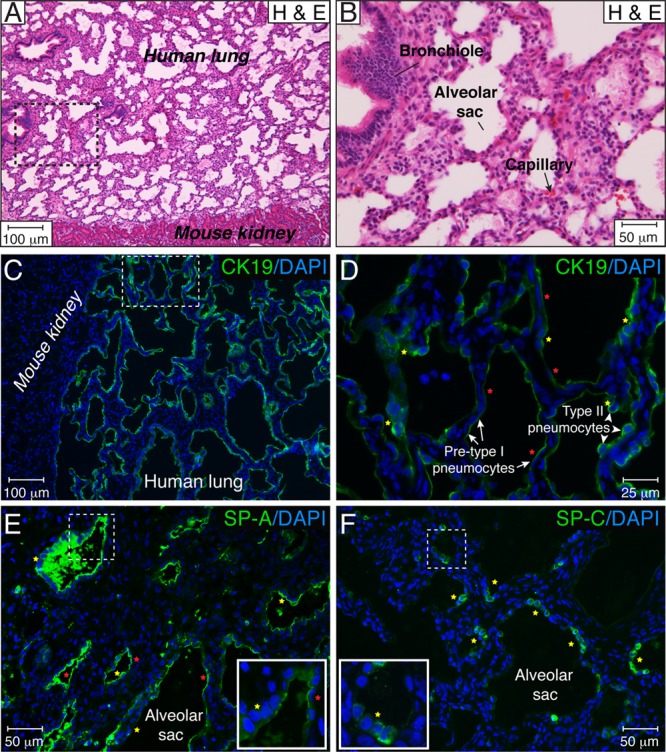
Extensive differentiation of human fetal lung tissue implanted under the kidney capsule of CB17-scid mice. (A and B) Fetal lung tissue implanted under the mouse kidney capsule developed over a period of 5 weeks. Histological tissue sections were stained with hematoxylin and eosin (H & E). The area outlined by the dotted lines in panel A is magnified in panel B. (C and D) The alveolar surface of the lung is formed by at least two types of epithelial cells, type II pneumocytes, with a cuboidal shape (yellow stars), and flattened, presumably pre-type I pneumocytes (red stars). Alveolar pneumocytes were visualized by CK19 immunostaining, and nuclei were counterstained with DAPI. The area outlined by the dotted lines in panel C is magnified in panel D. (E and F) During lung implant development, type II pneumocytes (yellow stars) robustly produced SP-A (E) and SP-C (F). Flattened, presumably pre-type I pneumocytes (red stars) were negative for SP-C (F), but their apical surface stained positive for SP-A (E), suggesting a premature phenotype of the type I cells. The dotted square shows the area of the inset. These results are representative of those observed for 10 implants each from three different lungs developed over 4 to 6 weeks.