
Fig 7.
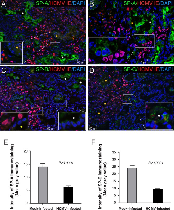
HCMV infection impaired surfactant protein expression in alveolar epithelial cells. (A to D) Histological sections of lung implants (cohort B) 7 days after virus inoculation were coimmunostained for HCMV IE and SP-A (A and B), SP-B (C), and SP-C (D). Nuclei were counterstained with DAPI. (A) Alveolar epithelial cells, identified by CK19 staining of the adjacent sections, had less SP-A secretion in the center of the viral lesion (inset, yellow star) but not in the periphery (white star). (B) Analysis of an alveolar duct at the edge of the viral lesion by laser confocal microscopy revealed diminished SP-A expression levels in pneumocytes proximal (yellow star) but not distal (white star) to the lesion. (C) SP-B was produced by the lung implant at a lower level than SP-A (inset, white star), and its levels further decreased in infected pneumocytes (inset, yellow star). (D) Type II pneumocytes expressing SP-C were not clearly evident in infected lung implants. Rare SP-C-positive type II pneumocytes (white star) were detected at the edge of the viral lesion but not in the center (yellow stars). (E and F) Representative data for SP-A (E) and SP-C (F) immunolabeling quantifications. Fourteen areas of the viral lesion from 1 implant and 14 areas from 3 mock-infected implants were quantified. Error bars represent standard errors of the means. As indicated by the fluorescence intensity, the expression levels of SP-A and SP-C were significantly reduced in HCMV-infected lung implants (P < 0.0001 by Mann-Whitney U test). These results are representative of those observed for 2 infected implants and 5 mock-infected implants obtained from two different lungs (cohorts A and B).