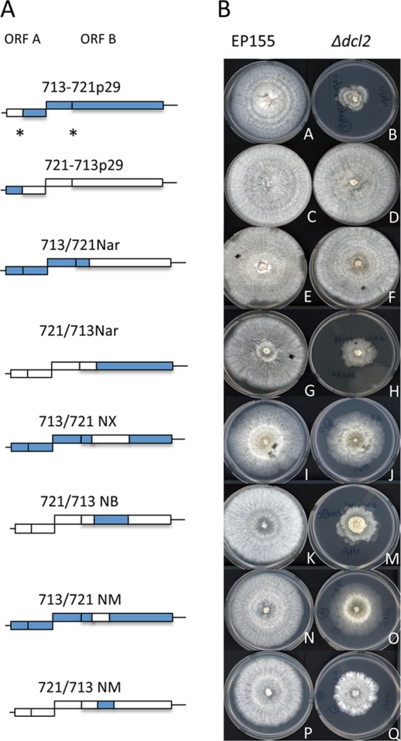
Fig 4.

Colony morphologies conferred by CHV-1/EP713–CHV-1/EP721 chimeric viruses in wild-type strain EP155 and the Δdcl2 mutant strain. (A) Schematic diagrams of chimeric viruses. The genomes of CHV-1/EP713 and CHV-1/EP721 contain two open reading frames designated ORF A and ORF B, as indicated at the top. The coding regions of individual chimeric viruses derived from the CHV-1/EP713 genome are indicated as blue boxes, and the CHV-1/EP721 genome-derived coding regions are indicated as white boxes. The autocatalytic cleavage sites for p29 and p48 are indicated by asterisks. The 5′ and 3′ noncoding terminal regions are indicated as lines. (B) Colony morphologies conferred by CHV-1/EP713–CHV-1/EP721 chimeric viruses in wild-type strain EP155 (left column) and the Δdcl2 mutant strain (right column). Infections were initiated by transfection with transcripts of the corresponding chimeric virus (3), and infected colonies were transferred to potato dextrose agar. The photographs were taken on day 7 of the experiment.
