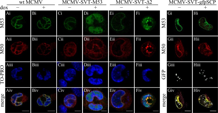
Fig 7.
Cellular localization of pM50 in the presence of mutant pM53. M2-10B4 cells were infected with wt MCMV (columns A and B); MCMV-SVT-M53, a virus expressing a second wt M53 allele (columns C and D); MCMV-SVT-Δ2 (columns E and F); and MCMV-SVT-gfpSCP (columns G and H) at an MOI of 0.5 in the absence (−) or presence (+) of 1 μg/ml doxycycline and fixed 36 h p.i. The cells were costained with polyclonal antisera specific for pM53 and pM50, followed by Alexa Fluor-conjugated secondary antibodies, and analyzed under a confocal immunofluorescence microscope. DNA was visualized using To-Pro 3. Scale bar, 10 μm.

