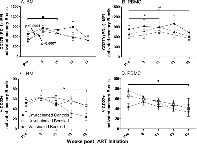
Fig 9.
CD279 (PD-1) and CD22 levels on activated memory B cells in BM and PBMC during the study. (A) Mean fluorescence intensity (MFI) of PD-1 on BM activated memory B cells. *, significant increase in expression levels over 11 weeks of ART (P = 0.0089 for unvaccinated boosted macaques; P = 0.0005 for vaccinated boosted macaques). (B) MFI of PD-1 on PBMC activated memory B cells. *, significant increase in expression levels over 11 weeks of ART (P = 0.028 for unvaccinated boosted macaques; P = 0.0070 for vaccinated boosted macaques); #, decline in PD-1 MFI at week 19 compared to pre-ART values for all groups combined (P = 0.029). (C) CD22 expression on activated memory B cells in bone marrow. *, significant negative slope of the vaccinated boosted macaque group over weeks 8 to 19 (P = 0.0021). (D) CD22 expression on activated memory B cells in PBMC. *, significant negative slopes of unvaccinated boosted and vaccinated boosted macaque groups from pre-ART values to week 19 (P = 0.017 and P < 0.0001, respectively). Plotted are group means ± standard errors of the means.

