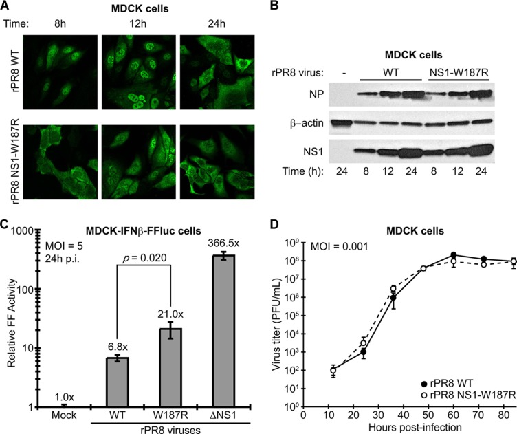
Fig 2.
Characterization of WT and NS1-W187R viruses in vitro. (A) NS1 localization during infection. Shown is indirect immunofluorescence analysis of NS1 protein localization in MDCK cells infected for the indicated times with rPR8 WT or rPR8 NS1-W187R viruses (multiplicity of infection [MOI] of 2 PFU/cell). The primary antibody was pAb 155 (12). (B) Expression of viral NS1 and NP proteins during infection. SDS-PAGE and Western blot analysis of lysates from MDCK cells infected for the indicated times with rPR8 WT or rPR8 NS1-W187R viruses (MOI of 5 PFU/cell). NS1 was detected using pAb 155, NP was detected using monoclonal antibody (MAb) HT103 (20), and β-actin was detected using MAb A4700 (Sigma-Aldrich, St. Louis, MO). (C) Induction of IFN-β by different rPR8 mutants. MDCK-IFN-β-FF-Luc cells were infected at an MOI of 5 PFU/cell for 24 h with the indicated virus (or mock infected) prior to analysis of luciferase activity. p.i., postinfection. Bars represent mean values (n = 3), and error bars represent standard deviations (SD). (D) Multicycle growth analysis of rPR8 WT and rPR8 NS1-W187R viruses in MDCK cells. Data points show mean values (n = 3), and error bars represent SD.

