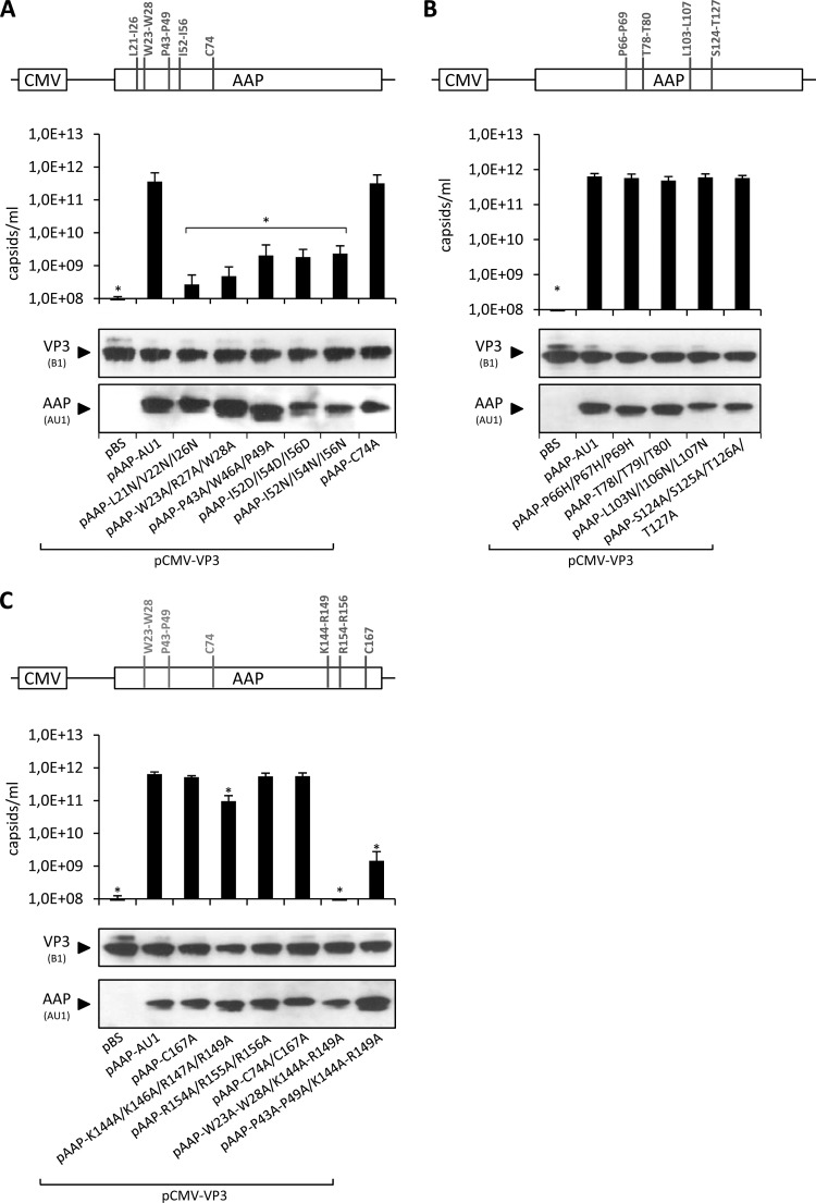
Fig 3.
Capsid assembly by amino acid exchange mutant AAPs. Amino acids at selected sequence motifs in the N-terminal (A), central (B) and C-terminal (C) parts of AAP were exchanged as schematically depicted. Capsid assembly was analyzed after cotransfection of 293T cells with the respective mutant AAPs or pAAP-AU1 together with capsid protein VP3 (pCMV-VP3) via A20 antibody-based capsid ELISA. Coexpression of VP3 with the empty pBS vector served as a negative control. Protein expression was analyzed by Western blot assay using MAb B1 (detection of VP3) and the AU1 tag MAb. Bars represent the average capsid titers of at least three independent experiments. Asterisks indicate capsid titers significantly lower than that obtained with pAAP-AU1 (P < 0.01).

