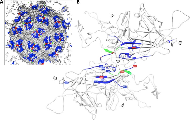
Fig 8.
Capsid protein domains contributing to AAV2 capsid assembly. (A) Schematic representation of the inner capsid surface of AAV2. Positions of capsid assembly defect mutant constructs described previously (3, 26) are shown in blue. Mutant constructs described in this study and by Popa-Wagner et al. (18) are shown in red, while the B1 antibody epitope is shown in green. Note the clustering of assembly mutant constructs at the 2-fold symmetry axes. (B) Ribbon drawing of a VP dimer highlighting the positions of assembly defect mutant constructs described previously (blue) and in this study (red). The B1 epitope is shown in green. The 3-fold and 5-fold axes of symmetry are indicated by triangles and pentamers, respectively.

