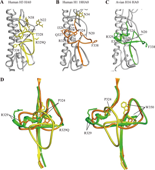
Fig 3.
Structural comparison of the H16HA0 cleavage site with other HA0s. HA2 domains for human H3HA0 and human H1 18HA0 were aligned with H16HA0. The cleavage sites are colored yellow for human H3HA0 (A), orange for human 18HA0 (B), and green for avian H16HA0 (C). (D) Overlay of the cleavage sites of H3HA0, 18HA0, and H16HA0. The two views differ by a rotation of 90° about the 3-fold vertical axis. For H3HA0, the cleavage site forms a loop structure that projects from the glycoprotein surface, while for 18HA0, the cleavage site forms a loop structure that abuts the glycoprotein surface. Extraordinarily, in H16HA0, the cleavage site forms an α-helix structure that has not been observed before.

