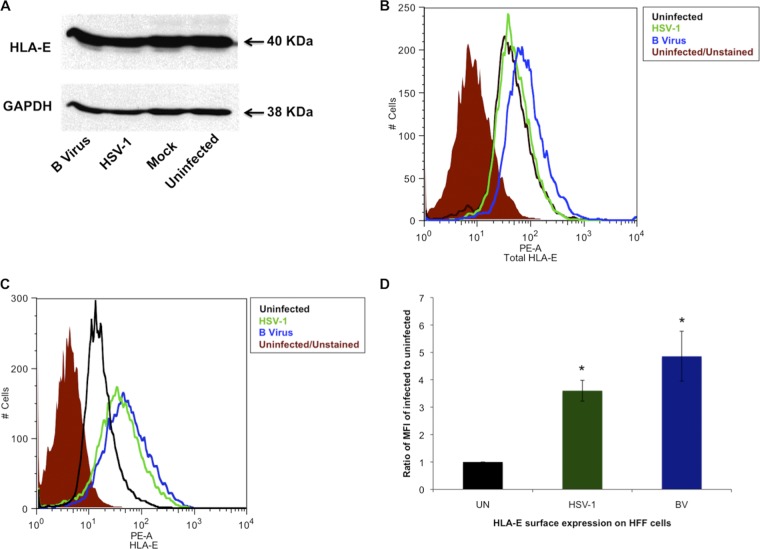
Fig 3.
Regulation of HLA-E in HFF cells infected with B virus. (A) Western blot analysis of HFF cells infected with B virus or HSV-1 and probed with anti-HLA-E polyclonal antibody. (B) HFF cells were infected with B virus or HSV-1 at an MOI of 10, and total HLA-E expression was determined by using flow cytometry. The shaded histogram represents uninfected cells unstained for autofluorescence, the black histogram represents uninfected cells stained with PE-conjugated anti-HLA-E antibody clone 3D12 (PE-A), the blue histogram represents B virus-infected stained cells, and the green histogram represents HSV-1-infected stained cells. (C) HLA-E surface expression determined by using flow cytometry. (D) MFIs of B virus (BV)- and HSV-1-infected HFF cells relative to the MFI of uninfected (UN) HFF cells. *, P ≤ 05.

