
Fig 6.
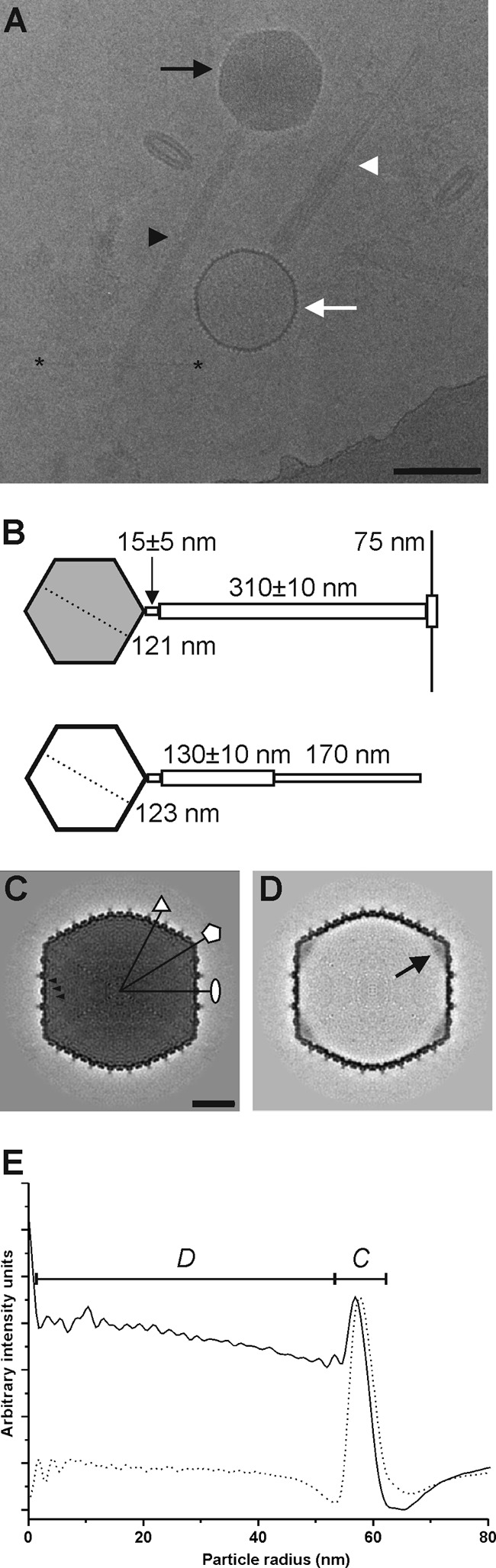
Organization of ϕR1-37. (A) Electron cryomicrograph (2.5 μm underfocus) of ϕR1-37 showing intact DNA-containing virions (black arrow) with extended tails (black arrowhead) and empty DNA-lacking particles (white arrow) with contracted tail sheaths (white arrowhead). The clear tail fibers are visible starting at the tip of the extended tail and reaching to the sides (the ends of two tail fibers are indicated with asterisks). Bar, 100 nm. (B) Schematic representation of an intact (gray head) and an empty (white head) ϕR1-37 particle with uncontracted and contracted tails, respectively. The numbers indicate the sizes obtained in this study. (C) A 0.45-nm-thick central section through the 23.8-Å-resolution DNA-containing virion reconstruction. The symmetry axes are indicated with a white ellipse (2-fold), triangle (3-fold), and pentagon (5-fold). The black arrowheads point to three successive layers of packaged DNA. Bar, 20 nm. (D) A 0.45-nm-thick central section through the 23.4-Å-resolution DNA-lacking particle reconstruction. A black arrow points to the connector density at one of the 5-fold vertices. (E) Radial density profiles of the icosahedral reconstructions of the DNA-containing (solid line) and DNA-lacking (dashed line) viral particles. The capsid density is indicated with a C, and the DNA with a D.