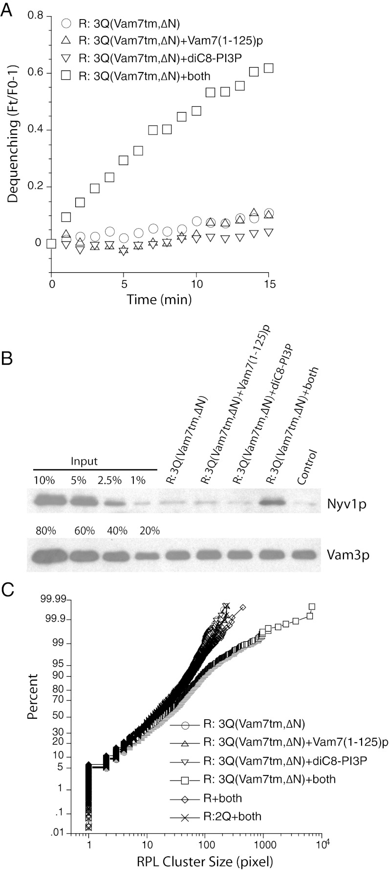
Fig. 5.
The Vam7 N-domain promotes trans-SNARE complex-dependent RPL docking. R-SNARE RPLs and 3Q-SNARE RPLs bearing Vam7tm,∆N were incubated at 27 °C for 15 min with Vam7(1-125)p from which the GST domain had been cleaved, diC8-PI3P (47 μM), or both. (A) NBD dequenching, expressed as its ratio over the fluorescent signal at t = 0. At the end of the 15-min incubation, samples were transferred to ice, solubilized in RIPA buffer, and subjected to immunoprecipitation using immobilized anti-Vam3p antibody (21). Western blotting of the immunoprecipitated Vam3p and the coprecipitated Nyv1p are shown in B. As control for protein interaction in membrane lysates, R-SNARE RPLs were incubated separately from 3Q-SNARE RPLs [which had Vam7(1-125)p and diC8-PI3P] and mixed immediately after the addition of RIPA buffer. (C) To measure the clustering of R-SNARE RPLs under various reaction conditions, 1 μL from each sample was diluted 40-fold in 20 mM Hepes-KOH, pH 7.5, 150 mM NaCl, and 10% glycerol. An aliquot (4 μL) of the diluted sample were transferred to a microscope plate and covered by a 22-mm2 coverslip. Samples were observed with an Olympus BX51 microscope with a 100-W mercury arc lamp, with images captured by a CCD camera. The size distribution was analyzed by ImageJ and plotted accordingly (26).

