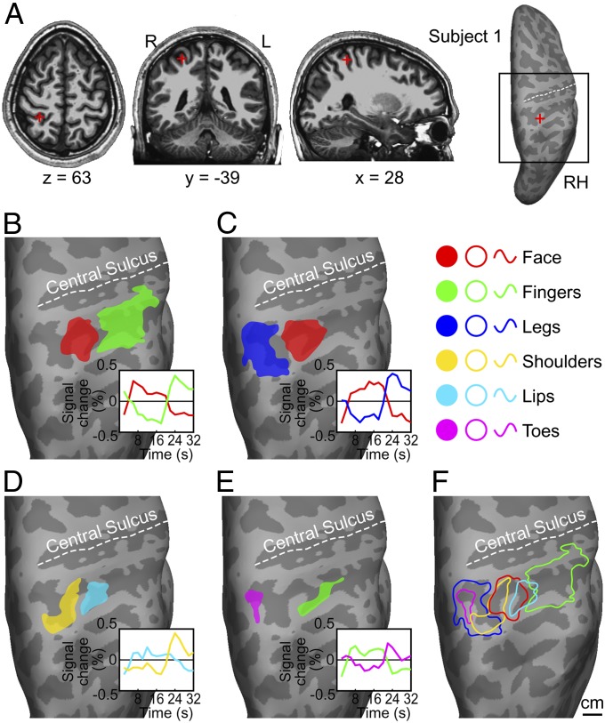
Fig. 2.
Parietal face and body areas in a representative subject. (A) Anatomical location and Talairach coordinates of the parietal face area in structural images (Left three panels) and on an inflated cortical surface (Rightmost panel). The black square indicates the location of a close-up view of the superior posterior parietal region shown below. RH, right hemisphere. (B–E) Body-part ROIs and their average signal changes (Insets) for (B) face vs. fingers scans, (C) face vs. legs scans, (D) lips vs. shoulders scans, and (E) fingers vs. toes scans. (F) A summary of parietal face and body areas. Contours were redrawn from the ROIs in B–E. To reduce visual clutter, contours of face and finger ROIs from C and E were not shown in F.

