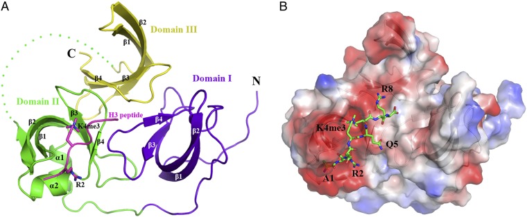
Fig. 1.
Overall structure of Spindlin1. (A) A ribbon diagram of the Spindlin1 tudor-like domains in complex with an H3K4me3 peptide. The three tudor-like domains of Spindlin1, following the order from the N-terminal to the C-terminal end, are colored blue, green, and yellow, respectively. The histone H3 peptide containing residues 1–8 with trimethylated K4 residue is shown in magenta. The trimethylated H3K4 sidechain is shown in a stick model. (B) Electrostatic potential on the surface of Spindlin1. The structure is viewed from the same direction as in A, and the peptide is shown in a stick representation (carbon, green; nitrogen, blue; oxygen, red).

