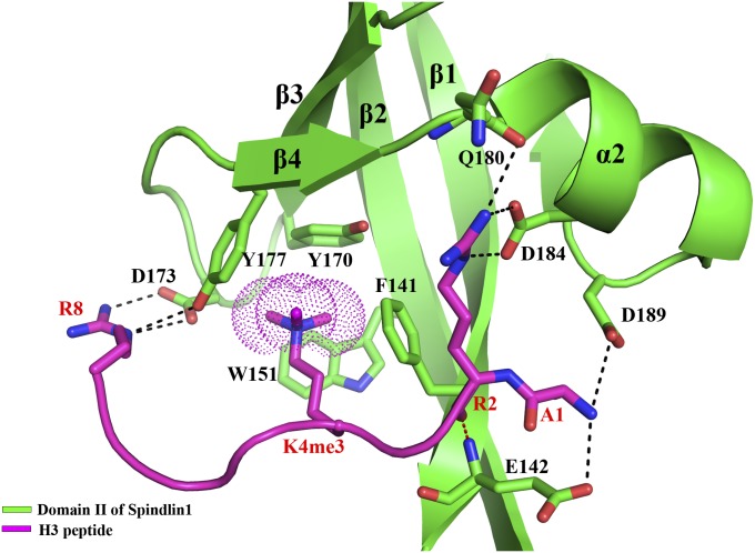
Fig. 2.
A close view of histone H3K4me3 peptide binding by domain II of Spindlin1. The Spindlin1 domain is shown in a ribbon representation colored green, and the residues involved in interaction with the histone peptide are superimposed as a stick model (carbon, green; nitrogen, blue; oxygen, red). The histone peptide is shown as a magenta coil, and the residues interacting with Spindlin1 are superimposed as a stick model (carbon, magenta). The trimethyl group of H3K4me3 is highlighted with dots. Dashed lines indicate intermolecular hydrogen bonds (red dashed lines indicate those between mainchain atoms).

