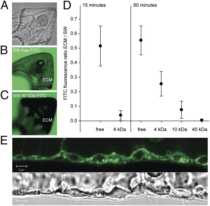
Fig. 2.
Determination of epithelial permeability in pluteus larvae in vivo using FITC dextran conjugates of varying molecular weights and confocal microscopy. (A) Transmission image of pluteus larva; (B) confocal image of larva in a similar position as in A exposed to 4-kDa FITC dextran in seawater for 60 min; the green color in the extracellular matrix (ECM) indicates equilibration of FITC dextran between surrounding seawater (SW) and ECM; (C) confocal image of larva exposed to 40 kDa for 60 min; the dark color indicates lack of equilibration of FITC dextran between ECM and SW. (D) Equilibration of FITC dextrans of varying molecular weights between SW and ECM following 15- and 60-min incubation. Equilibration is represented as the ratio of FITC fluorescence within the ECM and the SW surrounding the larva. Low values indicate a low permeability. (E) Corresponding confocal (FM1-43-stained) and transmission images of the pluteus larva outer epithelium. Bars represent ±SD; n = 9.

