Fig. 3.
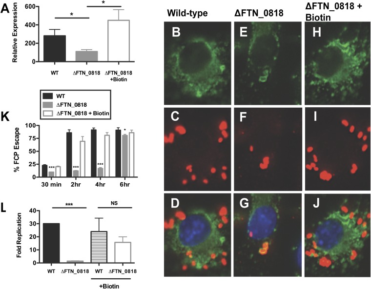
Biotin rescues rapid phagosomal escape and the ΔFTN_0818 replication defect in macrophages. (A–J) Macrophages were infected, and qRT-PCR was used to measure the expression of iglA and normalized to the expression of uvrD at 30 min pi (*P < 0.05) (A) and immunofluorescence microscopy was used to determine escape kinetics of WT (B–D), ΔFTN_0818 (E–G), and ΔFTN_0818 supplemented with biotin (H–J) 2 h pi (FITC-stained LAMP-1, green; anti-Francisella, red; DAPI, blue). (K) Two hundred bacteria were counted per sample, and colocalization with lysosomal-associated membrane protein 1 (LAMP-1) was used as a marker for phagosomal localization. *P < 0.05; ***P < 0.0001. (L) Macrophages were infected with WT or ΔFTN_0818 strains in media with or without biotin. Colony-forming units were quantified 30 min and 6 h pi, and fold replication was calculated.

