Fig. 3.
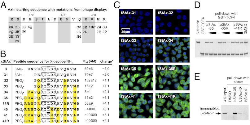
Cell permeability and β-catenin binding of StAx peptides. (A) Summary of variants observed at least twice in the 32 selected phage sequences. Numbers indicate the frequency of appearance at a position (complete list in Fig. S3B). (B) Stapled peptide sequences including their Kd (mean ± SE) with β- catenin and overall charge (calculated for fStAx at pH 7.5 with Marvin 5.2.3, 2009, ChemAxon). (C) Analysis of DLD1 cellular uptake of fStAx peptides (7.5 μM, 24 h) by confocal fluorescence microscopy (overlaid images with blue: nuclear DAPI, green: fluorescein; details in Fig. S5). (D) In vitro competition of aStAx peptides (0.1, 0.5, 2.5 μM) with bead-immobilized GST-TCF4(1-52) for β-catenin (0.5 μM). (E) Pull-down assay with bStAx peptides in DLD1 lysates.

