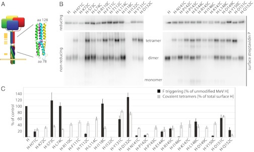
Fig. 6.
Membrane-distal (upper) H stalk sections maintain close physical proximity during F triggering. (A) Overview of the cysteine-scanning mutagenesis performed along the H stalk, represented by a yellow bar (Left) in the cartoon. (Inset) Homology model of the central section of the MeV H stalk, generated in Swiss-MODEL based on the coordinates of a PIV5 HN stalk fragment (PDB ID 3TSI). (B) Assessment of the covalent oligomerization status of cell-surface–exposed H cysteine mutants under reducing and nonreducing conditions. (C) Quantitation of relative amounts of surface-exposed H in covalent tetramers (based on densitometric analysis of gels as shown in B) and F-triggering activity; data represent average of four experiments ± SEM.

