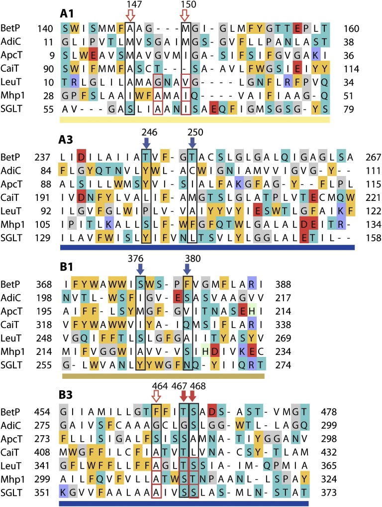
Fig. 2.
Structure alignment of regions involved in sodium binding in FIRL-fold transporters, i.e., helices A1, A3, B1, and B3, highlighting positions potentially forming the Na1′ site in BetP (blue arrows, black boxes) and the Na2 site in BetP (red arrows, black boxes). Filled arrows indicate side-chain interactions; open arrows indicate backbone interactions in BetP. Residues forming the Na2 binding site in other transporters are outlined with red boxes. Background colors indicate amino-acid types as follows: negatively charged (red), positively charged (blue), other polar (cyan), aromatic (gold), helix-breaker (gray), and histidines (pale green). Bars under each segment are colored according to Fig. 1.

