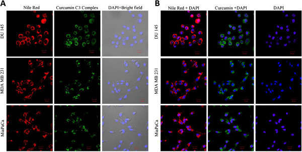
Figure 9.
Confocal images of different cancer cell lines incubated with PLGA-CURC - (A) Red :- nile red-labeled PLGA-CURC, Green:- curcumin, Bright field merged with cell nuclei stained with DAPI; (B) Merged images of nile red-labeled PLGA-CURC with DAPI; Merged images of curcumin with DAPI and DAPI.

