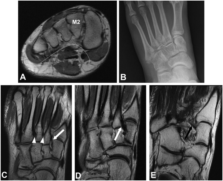
Figure 3.
Axial FSE at the level of a normal Lisfranc joint (A) demonstrates asymmetric arch configuration of the osseous structures, with the second metatarsal base (M2) as the keystone. Anteroposterior radiograph of the foot (B) demonstrates mortise configuration at the tarsometatarsal joint, as the second metatarsal base is recessed between the medial and lateral cuneiforms. Coronal FSE images of the midfoot (c-e) demonstrate the dorsal (white arrow) and plantar (black arrow) bands of the Lisfranc ligament, as well as the second and third intermetatarsal ligaments (white arrowhead).

