
Figure 2.
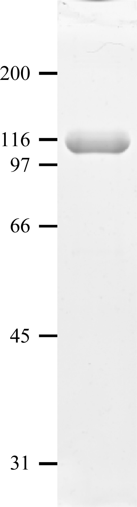
SDS–PAGE analysis. 5 µg purified CPIIIHis was analyzed on a 10% acrylamide gel under nonreducing conditions followed by Coomassie Blue staining. Migration positions of marker proteins are indicated in kDa.
Official websites use .gov
A
.gov website belongs to an official
government organization in the United States.
Secure .gov websites use HTTPS
A lock (
) or https:// means you've safely
connected to the .gov website. Share sensitive
information only on official, secure websites.
SDS–PAGE analysis. 5 µg purified CPIIIHis was analyzed on a 10% acrylamide gel under nonreducing conditions followed by Coomassie Blue staining. Migration positions of marker proteins are indicated in kDa.