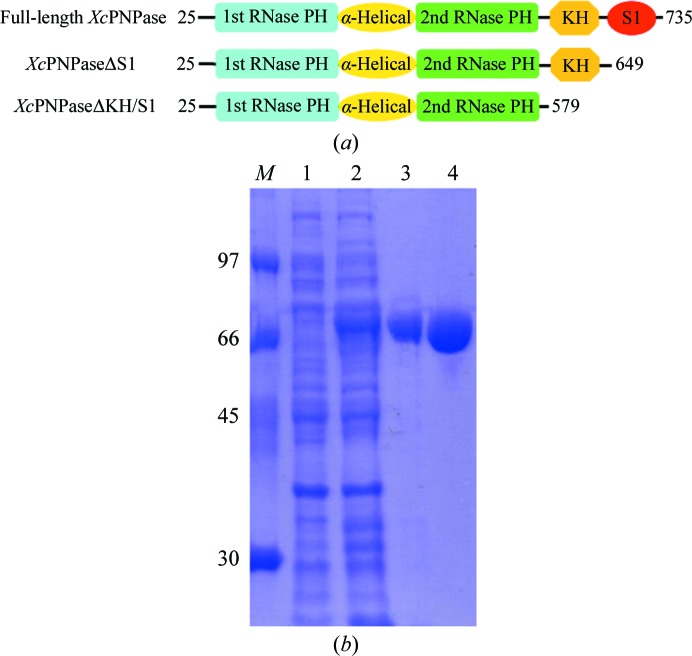
Figure 1.
(a) The domain architecture and constructs of XcPNPase used in these studies. The starting and ending residues are numbered. (b) SDS–PAGE (12%) monitoring of the overexpression and purification of XcPNPaseΔS1. Lane M, protein markers (labelled in kDa); lane 1, whole cell lysate before IPTG induction; lane 2, whole cell lysate after IPTG induction; lane 3, nickel-column-purified XcPNPaseΔS1 after IPTG induction; lane 4, nickel column-purified XcPNPaseΔS1 after TEV cleavage.

