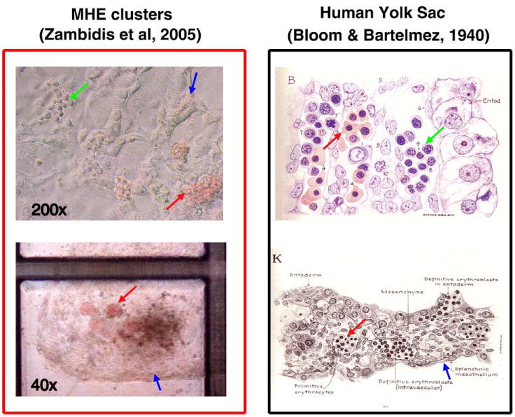
Figure 3.

Human ESC-derived MHE colonies possess many characteristics that resemble primary human yolk sac differentiation. Day 6 to 10 hEB cells recultured in serum-free methylcellulose containing hematopoietic growth factors can give rise to rare, unique MHE colonies (top left panel), which can be shown to consist of a mixture of mature CD31+VE-cadherin+ endothelial, vimentin+ mesenchymal, and primitive hematopoietic cells. If fed with fresh medium with growth factors, these clusters can become quite prolific after 5 to 6 weeks (bottom left panel). MHE clusters possess the following features that have great similarity to classic human yolk sac histologic architecture (right panels): (1) hematopoietic progenitors (green arrows) arise in intimate association with endothelium-mesenchymal-like cells (blue arrows); (2) the predominant hematopoietic progeny of both MHE colonies and yolk sac blood islands are nucleated primitive erythroblasts expressing embryonic/fetal hemoglobins (red arrows); and (3) primitive hematopoiesis precedes definitive erythro-myelopoiesis. Reprinted with permission from Bloom W, Bartelemez GW. Hematopoiesis in young human embryos.
