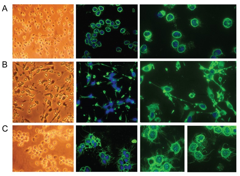
Figure 5. The C4B6− cells change morphology upon stimulation with various mitogens.
Left panels, inverted microscope pictures. Middle panels, fluorescence microscopy pictures of cells stained with anti-TO antiserum, captured with 40× objective. Right panels, fluorescence microscopy pictures of cells stained with anti-TO antiserum, captured with 63× objective. (A) Cells without mitogen, 24 hrs incubation. (B) Cells stimulated with Con A and PMA for 18 hrs. (C) Cells stimulated with LPS for 24 hrs.

