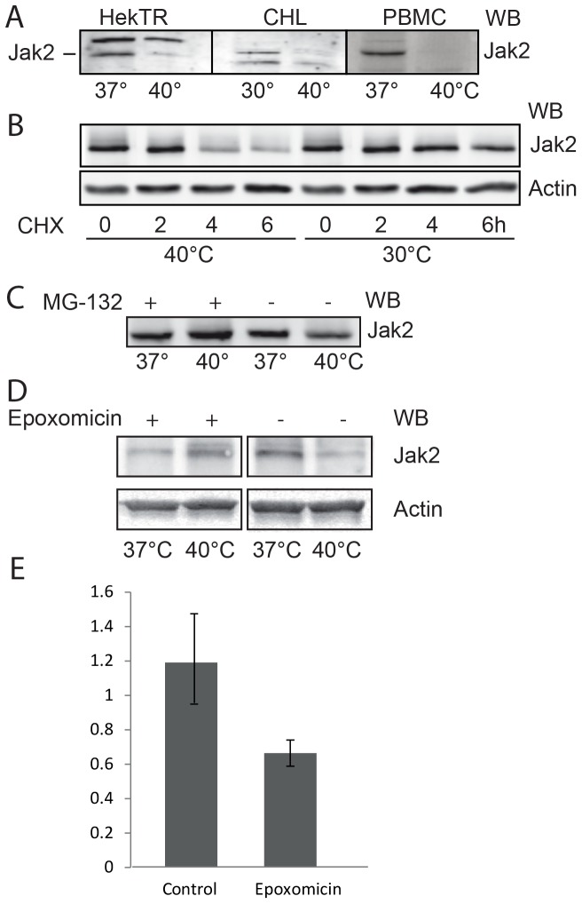
Figure 1. Effect of stressors on Jak2 protein levels in various cell types.
(A) Hek293-TR, Chinese hamster lung and PBM cells were incubated for 3 h, as indicated. The cells were lysed in 1% Triton X-100 and equal aliquots of the lysates were analyzed by western blotting (WB) for endogenous Jak2, using anti-Jak2 antibody. The antibody recognized a-specific bands in lysates of HekTR (above Jak2) and CHL (below Jak2). (B) In the presence of 20 µg/ml cycloheximide (CHX) Chinese hamster cells were transiently transfected with Jak2 and incubated at either 37 or 40°C. The cells were lysed in 1% SDS and equal aliquots were analyzed (WB) using anti-Jak2 and anti-actin antibodies. (C) Equal amounts of isolated PBMCs were incubated with or without 20 µM MG-132 at 37 or 40°C for 4 h. The cells were lysed in 1% SDS and equal amounts were analyzed (WB) using anti-Jak2 antibody. (D) Effect of Epoxomicin on Jak2 protein levels in PBMCs. Equal amounts of isolated PBMCs were incubated with or without10 µM Epoxomicin at 37 or 40°C for 4 h. The cells were lysed in 1% SDS and equal amounts were analyzed (WB) using anti-Jak2 antibody. The data in (A, B, C, D) are representative of three independent experiments. (E) Quantification of (D). The ratio of Jak2 to actin signals (western blot) were calculated. The data represent the mean of three independent experiments ± SEM.

