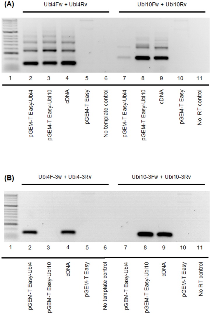
Figure 3. PCR amplification of Ubi4 and Ubi10 from various templates.
(A) Primers used for amplification of Ubi4 (Ubi4Fw and Ubi4Rv) and Ubi10 (Ubi10Fw and Ubi10Rv) are according to Hong et al. (2008) [5]. Lane 1: molecular weight marker (Bioline HyperLadder 1). The templates used are indicated for each lane. Empty vector (pGEM-T Easy) and no RT reactions were included as controls. The results shown are reverse gel images (dark bands on white backgrounds) from 25 amplification cycles. (B) Primers targeting the 3′-UTRs of Ubi4 (Ubi4-3Fw and Ubi4-3Rv) and Ubi10 (Ubi10-3Fw and Ubi10-3Rv) were used for amplification of the various templates. Lane assignments and templates used as in (A). Gel images are representative of 3 independent experiments.

