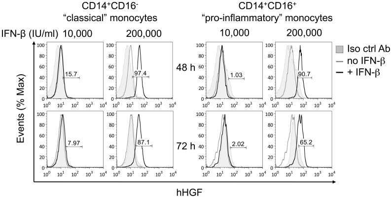
Figure 3. IFN–β increased cell–associated HGF in both human (A) CD14+CD16− “classical” and (B) CD14+CD16+ “pro-inflammatory” monocytes in a time– and-dose–dependent manner.
CD14+ monocytes were treated with IFN–β for the indicated time and dose, and cell–associated HGF was measured by flow cytometry. Cells were labeled with an antibody against anti–human HGF or isotype control antibody together with anti–CD14 and anti–CD16 antibodies. Histograms depict monoclonal anti–human HGF antibody (unfilled histograms) and isotype control antibody (filled histogram). Histograms depict representative data from two independent experiments. The values shown are the percentages of HGF positive cells as defined by fluorescence intensity greater than the control values of untreated cells.

