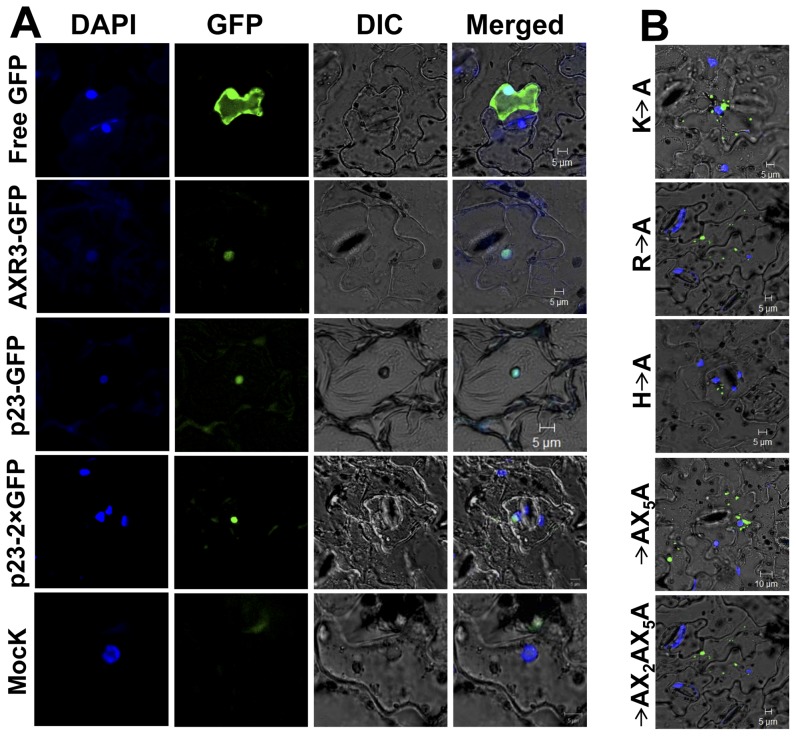
Figure 5. Nuclear localization of the p23 protein of HCRSV.
DAPI-stained nuclei (blue-color foci) were superimposed onto the differential interference contrast (DIC) image to form a merged image. (A) Kenaf leaves were infiltrated with free GFP and the signal was present in the entire cell including nucleus; p23-GFP and p23-2×GFP fusion proteins were only detected in the nucleus, similar to the positive control AXR3-GFP fusion protein. Free GFP represents agro-infiltration with vector lack of inserted gene. Mock represented agro-infiltration and no GFP signal was detected. (B) Localization of p23-GFP fusion protein to nucleus was affected by basic amino acid(s). Among the three basic amino acids (H, R and K) of p23 (20–41)-GFP protein, mutation in any one of the basic amino acids abolished nuclear localization of the p23-GFP fusion protein. Representative mutant images (H→A, R→A, K→A, RX5H→AX5A, KX2RX5H→AX2AX5A) were shown.

