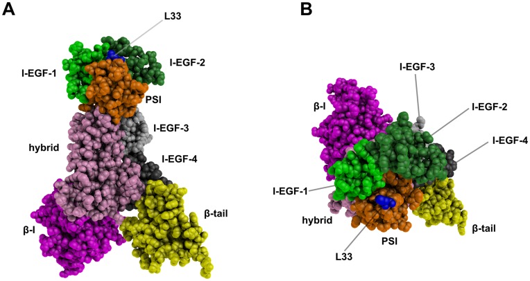
Figure 1. Ectodomain 3D structure model of the L33 β3 form.
A side view of β3 integrin (A) and an apical view of the β3 knee (B) are shown. Domains are differently colored and labeled and the L33 residue is shown in blue. These static views illustrate the HPA-1 polymorphic site that is located at the top of the β3 knee. These representations were generated using PyMOL software [16].

