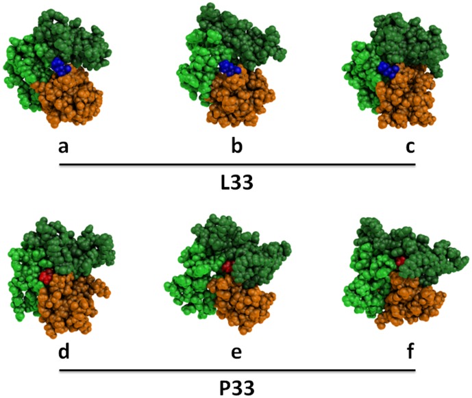
Figure 6. Snapshots of statistically representative structures of the PSI, I-EGF-1, and I-EGF-2 domains.
Three structure snapshots of L33 (a, b, and c) and of P33 (e, f, and d) β3 were chosen among the statistically most frequent structures in terms of rASA and number of contacts. L33 and P33 atoms are shown as blue and red spheres, respectively. All remaining atoms belonging to the PSI, I-EGF-1, and I-EGF-2 domains are shown as orange, light green and dark green spheres, respectively. In these snapshots, L33 is largely exposed at the β3 surface while P33 can be remote. P33 is in relatively close contact with the I-EGF-2 domain, but L33 is not. These representations were generated using PyMOL software [16].

