
Figure 1. Expression of ErbB receptor family members in human ovarian granulosa cell tumors (GCTs).
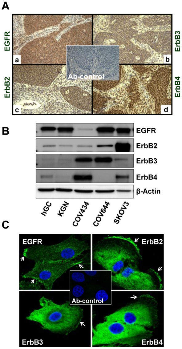
A) Immunohistochemistry was used to detect the expression of EGFR (a), ErbB2 (b), ErbB3 (c) and ErbB4 (d) in paraffin-embedded human GCT tissues. Magnification, 400×. B) Western blot detection of expression of ErbB family receptor proteins in human ovarian granulosa cells and GCT cell lines. SKOV-3 and COV644 cells were used as controls for the detection system. C) Fluorescent immunohistochemistry localization of ErbB receptors in KGN cells. The images were captured with a confocal laser scanning microscope. Arrows indicate the membrane localization of ErbBs in KGN cells. Magnification, 630×; Ab-control: 2nd antibody only control, magnification, 200×.