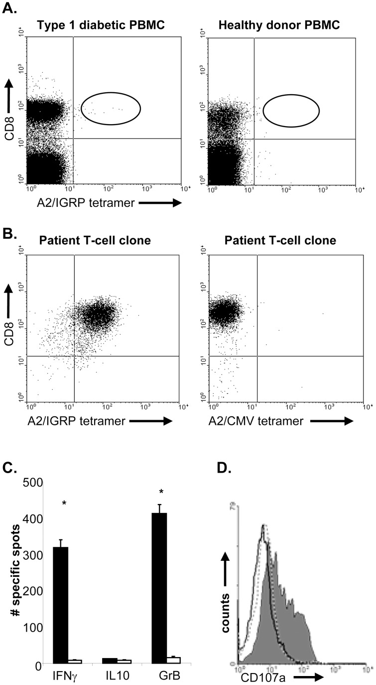
Figure 1. IGRP265–273-specific T-cells cloned from the peripheral blood of type 1 diabetic individuals.
A, PBMCs from a HLA-A*0201+ recent onset diabetic patients (left panel) and HLA A*0201+ healthy donors (right panel) were incubated with A2/IGRP tetramers, followed by incubation with anti-CD8. CD8/tetramer double positive T cells were only detected in PBMC obtained from type 1 diabetic individuals and were not detected in the blood obtained from healthy controls. B, CD8/tetramer double positive T-cells were sorted at one cell per well and clones were picked. IGRP-specific T-clones stained with IGRP specific tetramers were observed in wells derived from type 1 diabetic individuals (left panel). These clones did not bind control HLA-A2 tetramers (right panel). C, To assess their cytokine production profile, T-cells were incubated with IGRP peptide-pulsed or control peptide-pulsed HLA-A2 EBV-LCL on anti-IFNγ; anti-GrB and anti-IL10-coated ELISpot plates. Shown is the average number of spots of triplicate wells. Data are representative of 3 independent experiments. * indicates significant difference from controls, P<0.01. D, IGRP-specific T-cells were incubated with control peptide-pulsed (dashed line) or IGRP peptide-pulsed HLA-A2 EBV-LCL in the presence of anti-CD107a (grey histogram) antibodies. As a control, T-cells were incubated with IGRP peptide-pulsed target cells in the presence of isotype control antibodies (black line) for 5 hours. T-cells were stained for CD8 and expression of CD107a was analyzed on CD8+ T-cells using flow cytometry. Results are representative of 2 independent experiments.

