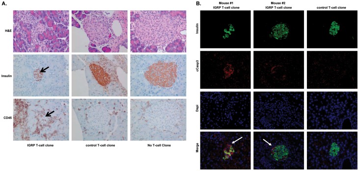
Figure 3. IGRP-specific T-cells are able to infiltrate and destroy beta-cells following intra-pancreatic injection into NOD-scidIL2rγnull HHD mice.
NOD-scid IL2rγnull HHD recipient mice were injected i.v. with 20×106 PBMC from an HLA-A*0201+ healthy donor. Two days later, these mice were injected intra-pancreatically with either 5×10⁶ IGRP-specific T-cells (left), 5×10⁶ control T-cells (middle) or were sham injected (right). Four weeks later, the pancreata were isolated and histologically examined. A, Sections of recipients of IGRP-specific or control T-cells, or of those receiving a sham injection were stained with H&E (upper panel) to visualize the histological integrity of the islets or stained for insulin (middle panel) to identify the beta cells or human CD45 (lower panel) to visualize the human T-cells. Similar data were obtained when staining sections for human CD8. B, Pancreatic sections were stained for insulin, cCaspase-3 to detect apoptotic cells and DAPI to identify nuclei. Individual fluorescence as well as an overlay is presented. Shown are representative examples of 5 mice per group.

