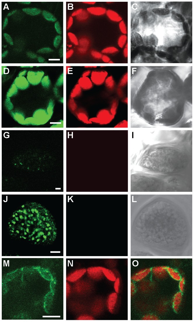
Figure 2. HvHMA1-GFP localizes to chloroplasts of leaves and intracellular compartments of aleurone layer cells of grains.

First row (A, B, C) shows a wild-type H. vulgare leaf cell, while the second row (D, E, F) shows a transgenic HvHMA1::GFP H. vulgare leaf cell. Third row (G, H, I) shows a wild-type aleurone layer cell and forth row (J, K, L) shows a transgenic H. vulgare aleurone layer cell expressing HvHMA1::GFP. Fifth row (M, N, O) shows a tobacco leaf cell transiently expressing HvHMA1::GFP under the control of the HvHMA1 promoter. A), D), G) J) and M) shows emission light at ∼525 nm. B), E), H) K) and N) shows chlorophyll autofluorescence. C), F), I) and L) transmission light images and O) is an overlay of M) and N). Bar is 5 µm.
